["7e2a2f2b3b32a29179d959013e1a74ab0cb7f604","d47466d1294abb83933ea7ddb79b56d460b55e40","807b351e9093860c9cb8d8da114b75f3ac09990c"]
Three-dimensional imaging of the breast
3D imaging
breast surgery
breast assessment
symmetry index
breast volume
3443
3443
Chapter reads
0
0
Chapter likes
7/10
Evidence score
07
7
Images included
03
3
Videos included
01
3D Imaging devices
3D Imaging devices
Due to technological advancements in the last two decades, imaging systems have become user-oriented and broadly available to plastic surgeons. Devices for 3D imaging include linear laser scanners such as the Minolta Vivid 910® (Konica Minolta Co. Ltd., Osaka, Japan) 123. The device uses the laser triangulation process, to create a 3D object 4.Two scanning systems used at our institution are stereophotogrammetry-based and structured-light-based systems. These are portable systems that are enabled for intraoperative use (Figure 1) 5.
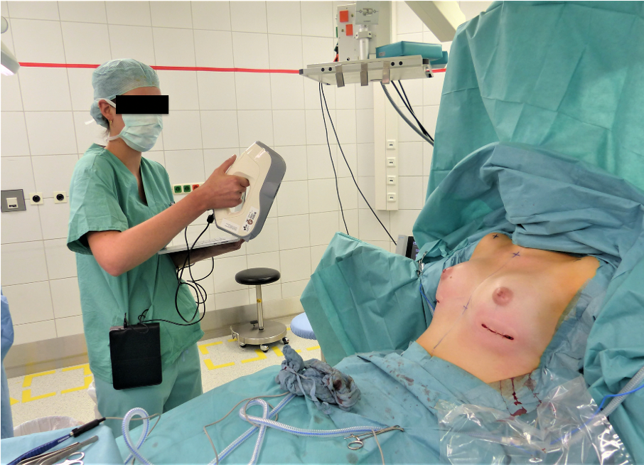
Figure 1 Intraoperative usage of the hand-held structured light scanner Artec EVA® (Artec 3D, Luxembourg).
Structured-light imaging uses a light pattern that is projected on the target to translate the targets surface into 3D coordinates 6. A device used at our institution is the Artec EVA® (Artec 3D, Luxembourg). It is a hand-held 3D scanner that has a reported accuracy of 0.5mm when reproducing the patient’s surface information 6. It has been validated for assessing breast morphology and shape 758. The scanner can be used intraoperatively, allowing advancement in the field of intraoperative breast assessment9 . Figure 1 shows the intraoperative usage of the EVA. Stereophotogrammetry uses multiple pictures and matches the intersections between different images into a 3D breast model 10. At our institution the Vectra H2® (Canfield Scientific, Inc., USA) is used. For breast assessment, a frontal 180º 3D model is generated using the Vectra Breast Sculptor® (Canfield Scientific, Inc., USA). Figure 2 shows the usage of the Vectra H2.
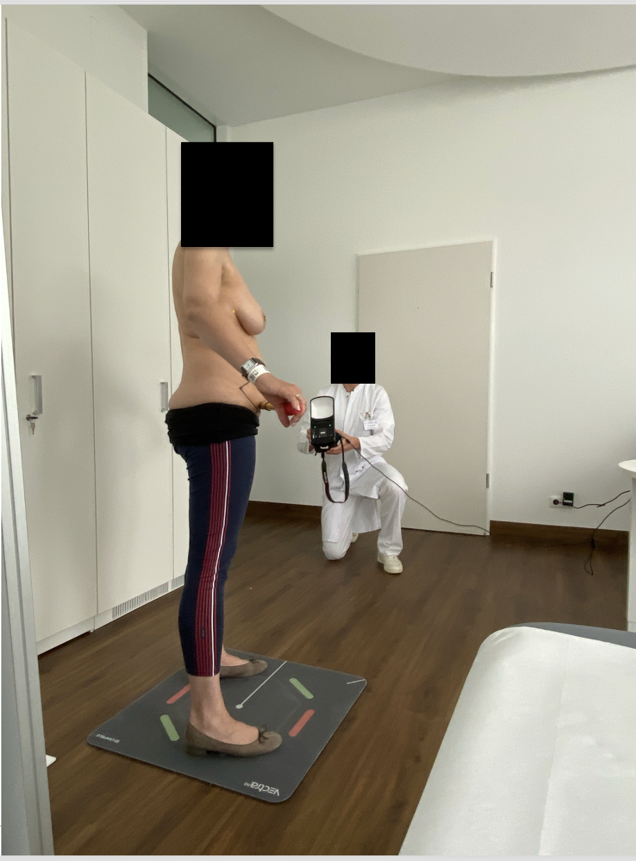
Figure 2 Use of the handheld stereophotogrammetry device Vectra H2® (Canfield Scientific, Inc., USA).
02
3D Imaging software
3D Imaging Software
Modern 3D scanners include custom-made software for point cloud processing and mesh generation. Recent devices offer standard evaluation- and simulation-tools. Canfield devices, such as the Vectra H2®, use the custom-made software Vectra Breast Sculptor® (Canfield Scientific, USA) and Vectra VAM® (Canfield Scientific, USA). The software allows outcome simulations as well as anthropometric and volumetric measurements of the breast region. The standard breast assessment that is performed by the Vectra is shown in Figure 3.
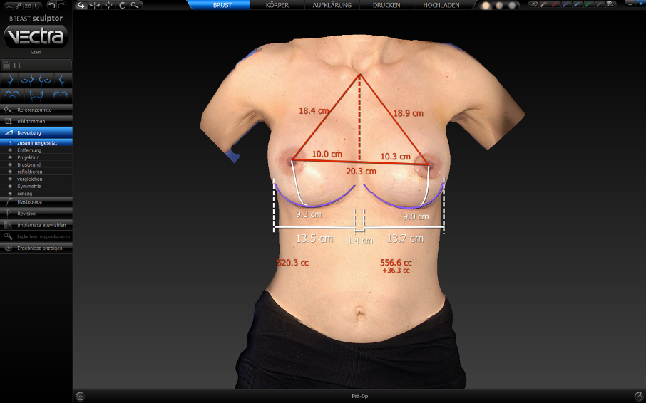
Figure 3 Vectra Breast Sculpture® (Canfield Scientific, USA); Apperance of a 43year/old women 8 days after silicone implant change using 385cc anatomical implants
Artec devices use the Artec Studio (Artec 3D, Luxembourg). In contrast to the Vectra Breast-Sculptor®, measurements are performed semi-automatically by selecting vertices on the mesh. Scientific approaches to sufficient digital breast assessment include the use of computer-aided design (CAD) software. Kovacs et al. used the Vivid 910®, (Konica-Minolta Co., Ltd., Osaka, Japan) and Raindrop Geomagic Studio® (Raindrop Geomagic, Inc., North Carolina, USA) to measure breast volume 2. The program showed good results in breast assessment. Other methods involve development of software for the use of 3D-models in experimental medicine. Georgii et al. presented a program which presents a software prototype for breast augmentation simulation 11. Earlier, we have presented a prototype software that uses all the common types of 3D file formats for digital anthropometry 12. The pre- and post-processing procedures of the 3D model are performed according to a standardized protocol. The prototyped graphical user interface (GUI) of this software is shown in Figure 4.
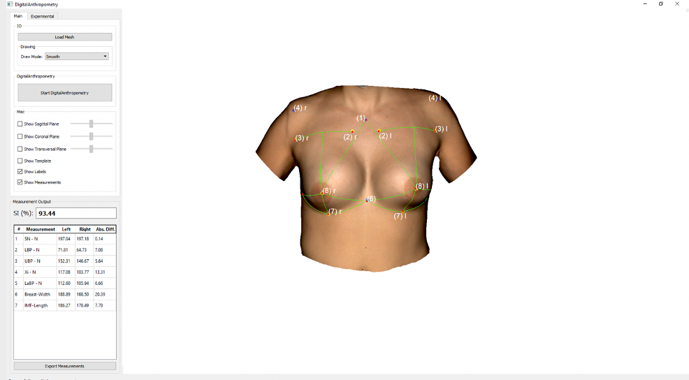
Figure 4 Our prototyped graphical user interface (GUI) of this software is shown Automatic measurements. Appearance of a 32-year-old patient twelve years after CBS using bilateral permanent silicone gel breast implant 225cc R/ 200cc L; mastopexy R+L; frontal view.
03
Digital assessment of the breast
Digital assessment of the breast region
To obtain comparable results, standardized protocols for digital breast assessment are required. Numerous protocols have been outlined for the use of 3D models for sufficient breast assessment13 14. A majority of these showed excellent results in breast surgery.
Landmarks
To assure adequate evaluation of the breast region, landmarks must be defined. The objective of landmark definition is to allow an approximation of the anatomical breast border. For analogous landmark definition, Brown et al. define seven key landmarks of a normal female breast: the suprasternal notch, the xiphoid, the medial end of the inframammary crease (IMF), the lateral end of the IMF, the lowest point of the IMF, the base of the breast, and the nipple15.
Eder et. al. defined a reproducible 3D breast symmetry evaluation protocol using eight landmarks13 . The improved protocol developed by our institution uses nine landmarks16 . These were chosen in concordance with the opinions of board-certified plastic surgeons. The process of landmark detection prior to scanning is shown in Video 1..
Eder et. al. defined a reproducible 3D breast symmetry evaluation protocol using eight landmarks13 . The improved protocol developed by our institution uses nine landmarks16 . These were chosen in concordance with the opinions of board-certified plastic surgeons. The process of landmark detection prior to scanning is shown in Video 1..
The following landmarks are marked using circular stickers (Figure 5):
(1) Sternal notch (SN) (2) Medial upper breast pole (MUBP)
(3) Lateral upper breast pole (LUBP)
(4) Coracoid process (CP)
(5) Lateral Breast Pole (LaBP)
(6) Xiphoid (Xi)
(7) Lower breast pole (LBP)
(8) Nipple (N)
(1) Sternal notch (SN) (2) Medial upper breast pole (MUBP)
(3) Lateral upper breast pole (LUBP)
(4) Coracoid process (CP)
(5) Lateral Breast Pole (LaBP)
(6) Xiphoid (Xi)
(7) Lower breast pole (LBP)
(8) Nipple (N)
Figure 5 Landmarks (1)-(8) (1) Sternal Notch (SN), (2) Medial Upper Breast Pole (MUBP), (3) Lateral Upper Breast Pole (LUBP), (4) Coracoid process (CP), (5) Lateral Breast Pole (LaBP), (6) Xiphoid (Xi), (7) Lower Breast Pole (LBP) and (8) Nipple (N).
Landmark (9), the Upper breast pole (UBP), is a guideline through landmarks (2) and (3) and must not be marked on the patient for digital breast assessment. The prototype software determines landmark (9) automatically.
Anthropometric measurements
Anthropometry of the breast is a commonly used technique in breast assessment that ensures adequate outcome evaluation. Smith et al. used six anthropometric measurements for breast assessment17 . These measurements consisted of the following distances: from lateral breast crease to nipple; axilla to nipple; nipple to midline; IMF to nipple; the lowest point of the breast to the nipple; nipple to most dependent point; and nipple to nipple.
Malata et al. describe a technique using four measurements: distance from nipple to sternal notch; distance from nipple to midline; angle subtended by the nipple-sternal notch line and the midline; and distance from center of nipple to IMF18 .
Malata et al. describe a technique using four measurements: distance from nipple to sternal notch; distance from nipple to midline; angle subtended by the nipple-sternal notch line and the midline; and distance from center of nipple to IMF18 .
The protocol developed by our institution uses seven anthropometric measurements (Figure 6):
(1) Sternal notch to nipple distance
(2) IMF to nipple distance
(3) Upper breast to nipple distance
(4) Xiphoid to nipple distance
(5) LaBP to nipple distance
(6) Breast width
(7) IMF length
(1) Sternal notch to nipple distance
(2) IMF to nipple distance
(3) Upper breast to nipple distance
(4) Xiphoid to nipple distance
(5) LaBP to nipple distance
(6) Breast width
(7) IMF length
These are measured between the landmarks defined by the stickers. The measurements are performed automatically by determining the geodesic distances between the landmarks using our prototype software. The prototyped GUI of the software is shown in Video 2.
Volumetric measurements
One of the most important features in 3D breast assessment are the volumetric measurements. A large number of analogous methods have been used to determine breast volume 19. The use of 3D imaging showed good results in breast volume assessment. Numerous studies have dealt with their validation. Eder et al. stated that 3D scanning and 3D imaging-supported volume determination is precise 20. Howes et al. confirmed that the precision of the laser-assisted 3D scan is equivalent to the MRI in volume determination 21. Novel state-of-the-art systems, such as the Vectra H2, can easily define breast volume (Video 3).
However, determining accurate breast volume still presents a major challenge in 3D imaging as the imaging systems generate surface information only. Studies conducted on scanner accuracy conclude that further research must be conducted on breast volume determination22 .
Outcome evaluation tools
To guarantee sufficient breast analysis, various outcome evaluation tools have been developed. Most methods described combine several parameters into one indicative value using experimental software.Fitzal et al. presented the breast assessment tool (BAT), an application for automatic symmetry quantification. It determined the breast symmetry index (BSI) using 2D photography and compared the areas and the circumference of a breast both breasts 23.
Cardoso et al. presented a piece of software for assessing the outcomes of patients who underwent breast cancer conservative treatment (surgery and radiotherapy) called the BCCT.core program 24. The aesthetic results of patients after BCT were automatically determined by 2D photography. Oliveira et al. adapted the method to a 3D imaging system 25. Mikolajczyk et al. presented the BreastIdea tool, a web application that uses 2D anthropometric data to analyze the effects of symmetry26 .The method proposed by our institution uses automatized digital anthropometry of 3D data to evaluate outcomes in breast surgery. We have presented a prototype software that combines digital anthropometric measurements into one value, the symmetry index (SI) 12. Earlier, we have used seven measurements for breast assessment12 .
Cardoso et al. presented a piece of software for assessing the outcomes of patients who underwent breast cancer conservative treatment (surgery and radiotherapy) called the BCCT.core program 24. The aesthetic results of patients after BCT were automatically determined by 2D photography. Oliveira et al. adapted the method to a 3D imaging system 25. Mikolajczyk et al. presented the BreastIdea tool, a web application that uses 2D anthropometric data to analyze the effects of symmetry26 .The method proposed by our institution uses automatized digital anthropometry of 3D data to evaluate outcomes in breast surgery. We have presented a prototype software that combines digital anthropometric measurements into one value, the symmetry index (SI) 12. Earlier, we have used seven measurements for breast assessment12 .
Examination sheet for 3D imaging of the breast region
Examination sheet for 3D imaging of the breast region
04
Limitations
Limitations
To this day, the use of 3D surface imaging has been undisputed. However, despite the numerous advantages of 3D surface imaging in breast assessment, it also has some limitations.One of the key aspects of using 3D imaging for clinical and scientific purposes is determining the method’s accuracy. Wesselius et al. compared three common imaging devices based on the accuracies of the devices. They concluded that the usefulness of absolute breast volume calculations from 3D surface images seems limited 22.In contrast, Eder et al. were able to determine resectionvolume in patients after mammareduction accurately using 3D imaging 20. Howes et al. confirmed in 2017 that the precision of the laser-assisted 3D scan is equivalent to that of the MRI in volume determination 21. Roostaeian and Adams determined the degree to which a preoperative simulation matches the actual postoperative result using Vectra M3 imaging 27. They concluded that 3D imaging can accuartly predicte breast volume27.
More research needs to be conducted on the accuracy of 3D imaging of the breast region. Technological advancements over the past two decades in 3D imaging simplified the creation of 3D models. However, scientific examinations of the breast region still require a trained user to guarantee inter-rater reliability. Therefore, the common software should not be used for scientific purposes without a standardized protocol. The protocol outlined by our institution presents a foundation for the assessment of the breast region using 3D imaging. Nevertheless, future studies may improve the protocol, which brings us one step closer to the long-term goal of establishing robust instruments to evaluate breast surgery results.
More research needs to be conducted on the accuracy of 3D imaging of the breast region. Technological advancements over the past two decades in 3D imaging simplified the creation of 3D models. However, scientific examinations of the breast region still require a trained user to guarantee inter-rater reliability. Therefore, the common software should not be used for scientific purposes without a standardized protocol. The protocol outlined by our institution presents a foundation for the assessment of the breast region using 3D imaging. Nevertheless, future studies may improve the protocol, which brings us one step closer to the long-term goal of establishing robust instruments to evaluate breast surgery results.
05
Conclusion
5. Conclusion
3D imaging of the breast region is an innovative tool for plastic surgeons and can support surgeons in their clinical everyday life. Plastic surgery is leading at applying this technology. While 3D imaging technology is improving, it enables surgeons to advance examination principles.
Images
Examination sheet for 3D imaging of the breast region
Figure 2 Use of the handheld stereophotogrammetry device Vectra H2® (Canfield Scientific, Inc., USA).
Figure 1 Intraoperative usage of the hand-held structured light scanner Artec EVA® (Artec 3D, Luxembourg).
Figure 3 Vectra Breast Sculpture® (Canfield Scientific, USA); Apperance of a 43year/old women 8 days after silicone implant change using 385cc anatomical implants
Figure 4 Our prototyped graphical user interface (GUI) of this software is shown Automatic measurements. Appearance of a 32-year-old patient twelve years after CBS using bilateral permanent silicone gel breast implant 225cc R/ 200cc L; mastopexy R+L; frontal view.
Figure 5 Landmarks (1)-(8) (1) Sternal Notch (SN), (2) Medial Upper Breast Pole (MUBP), (3) Lateral Upper Breast Pole (LUBP), (4) Coracoid process (CP), (5) Lateral Breast Pole (LaBP), (6) Xiphoid (Xi), (7) Lower Breast Pole (LBP) and (8) Nipple (N).
Figure 6 Anthropometric measurements
Videos
Video 2 The protocol developed by our institution for automated anthropometric measurements..
Video 3 The Vectra Breast-Sculptor® (Canfield Scientific, USA).
Video 1 The proposed protocol for landmark detection.
References
[1]
Optimization of 3-Dimensional Imaging of the Breast Region With 3-Dimensional Laser Scanners, Kovacs Laszlo, Yassouridis Alexander, Zimmermann Alexander, Brockmann Gernot, W??hnl Antonia, Blaschke Matthias, Eder Maximilian, Schwenzer-Zimmerer Katja, Rosenberg Robert, Papadopulos Nikolaos A., Biemer Edgar, Ovid Technologies (Wolters Kluwer Health), 2006
[2]
New Aspects of Breast Volume Measurement Using 3-Dimensional Surface Imaging, Kovacs Laszlo, Eder Maximilian, Hollweck Regina, Zimmermann Alexander, Settles Markus, Schneider Armin, Udosic Kristian, Schwenzer-Zimmerer Katja, Papadopulos Nikolaos A., Biemer Edgar, Ovid Technologies (Wolters Kluwer Health), 2006
[3]
Comparison between breast volume measurement using 3D surface imaging and classical techniques, Kovacs Laszlo, Eder Maximilian, Hollweck Regina, Zimmermann Alexander, Settles Markus, Schneider Armin, Endlich Matthias, Mueller Andreas, Schwenzer-Zimmerer Katja, Papadopulos Nikolaos A., Biemer Edgar, Elsevier BV, 2007
[4]
Review of 20 years of range sensor development, Blais Franc¸ois, SPIE-Intl Soc Optical Eng, 2004
[5]
Auf dem Weg zur objektiven Evaluation von Form, Volumen und Symmetrie in der Plastischen Chirurgie mittels intraoperativer 3D Scans, Koban K., Schenck T., Metz P., Volkmer E., Haertnagl F., Titze V., Giunta R., Georg Thieme Verlag KG,
[6]
Evaluation of the accuracy of a mobile and a stationary system for three-dimensional facial scanning, Modabber Ali, Peters Florian, Kniha Kristian, Goloborodko Evgeny, Ghassemi Alireza, Lethaus Bernd, Hölzle Frank, Möhlhenrich Stephan Christian, Elsevier BV, 2016
[7]
Evaluation of the accuracy of a mobile and a stationary system for three-dimensional facial scanning, Modabber Ali, Peters Florian, Kniha Kristian, Goloborodko Evgeny, Ghassemi Alireza, Lethaus Bernd, Hölzle Frank, Möhlhenrich Stephan Christian, Elsevier BV, 2016
[8]
Preoperative breast volume evaluation of one-stage immediate breast reconstruction using three-dimensional surface imaging and a printed mold, Chen Keng, Feng Chin-Jung, Ma Hsu, Hsiao Fu-Yin, Tseng Ling-Ming, Tsai Yi-Fang, Lin Yen-Shu, Huang Li-Ying, Yu Wen-Chan, Perng Cherng-Kang, Ovid Technologies (Wolters Kluwer Health), 2019
[9]
Auf dem Weg zur objektiven Evaluation von Form, Volumen und Symmetrie in der Plastischen Chirurgie mittels intraoperativer 3D Scans, Koban K., Schenck T., Metz P., Volkmer E., Haertnagl F., Titze V., Giunta R., Georg Thieme Verlag KG,
[10]
Three-dimensional Assessment of the Breast: Validation of a Novel, Simple and Inexpensive Scanning Process, ORANGES CARLO M., MADDURI SRINIVAS, BRANTNER PHILIPP, MSALLEM BILAL, GIORDANO SALVATORE, BENITEZ BENITO, KALBERMATTEN DANIEL F., SCHAEFER DIRK J., THIERINGER FLORIAN M., Anticancer Research USA Inc., 2019
[11]
“Topographic Shift”: a new digital approach to evaluating topographic changes of the female breast, Lotter Luisa, Brébant Vanessa, Eigenberger Andreas, Hartmann Robin, Mueller Karolina, Baringer Magnus, Prantl Lukas, Schiltz Daniel, Springer Science and Business Media LLC, 2021
[12]
Validation of a passive stereophotogrammetry system for imaging of the breast: A geometric analysis, Catherwood T., McCaughan E., Greer E., Spence R.A.J., McIntosh S.A., Winder R.J., Elsevier BV, 2011
[13]
New Aspects of Breast Volume Measurement Using 3-Dimensional Surface Imaging, Kovacs Laszlo, Eder Maximilian, Hollweck Regina, Zimmermann Alexander, Settles Markus, Schneider Armin, Udosic Kristian, Schwenzer-Zimmerer Katja, Papadopulos Nikolaos A., Biemer Edgar, Ovid Technologies (Wolters Kluwer Health), 2006
[14]
A Computational Tool for Preoperative Breast Augmentation Planning in Aesthetic Plastic Surgery, Georgii Joachim, Eder Maximilian, Burger Kai, Klotz Sebastian, Ferstl Florian, Kovacs Laszlo, Westermann Rudiger, Institute of Electrical and Electronics Engineers (IEEE), 2014
[15]
A Novel Method of Outcome Assessment in Breast Reconstruction Surgery: Comparison of Autologous and Alloplastic Techniques Using Three-Dimensional Surface Imaging, Hartmann Robin, Weiherer Maximilian, Schiltz Daniel, Seitz Stephan, Lotter Luisa, Anker Alexandra, Palm Christoph, Prantl Lukas, Brébant Vanessa, Springer Science and Business Media LLC, 2020
[16]
Objective breast symmetry evaluation using 3-D surface imaging, Eder Maximilian, Waldenfels Fee v., Swobodnik Alexandra, Klöppel Markus, Pape Ann-Kathrin, Schuster Tibor, Raith Stefan, Kitzler Elena, Papadopulos Nikolaos A., Machens Hans-Günther, Kovacs Laszlo, Elsevier BV, 2012
[17]
The Role of Three-Dimensional Scanning Technique in Evaluation of Breast Asymmetry in Breast Augmentation: A 100-Case Study, Liu Chunjun, Luan Jie, Mu Lanhua, Ji Kai, Ovid Technologies (Wolters Kluwer Health), 2010
[18]
A method of assessing female breast morphometry and its clinical application, Brown T.P.La H., Ringrose C., Hyland R.E., Cole A.A., Brotherston T.M., Elsevier BV, 1999
[19]
Objective breast symmetry evaluation using 3-D surface imaging, Eder Maximilian, Waldenfels Fee v., Swobodnik Alexandra, Klöppel Markus, Pape Ann-Kathrin, Schuster Tibor, Raith Stefan, Kitzler Elena, Papadopulos Nikolaos A., Machens Hans-Günther, Kovacs Laszlo, Elsevier BV, 2012
[20]
New aspects in digital breast assessment: further refinement of a method for automated digital anthropometry, Hartmann Robin, Weiherer Maximilian, Schiltz Daniel, Baringer Magnus, Noisser Vivien, Hösl Vanessa, Eigenberger Andreas, Seitz Stephan, Palm Christoph, Prantl Lukas, Brébant Vanessa, Springer Science and Business Media LLC, 2021
[21]
Breast Volume and Anthropomorphic Measurements, Smith David J., Palin William E., Katch Victor L., Bennett James E., Ovid Technologies (Wolters Kluwer Health), 1986
[22]
Congenital breast asymmetry: subjective and objective assessment, Malata C.M., Boot J.C., Bradbury E.T., Ramli A.R.B., Sharpe D.T., Elsevier BV, 1994
[23]
Objective Breast Volume, Shape and Surface Area Assessment: A Systematic Review of Breast Measurement Methods, Xi Wenjing, Perdanasari Aurelia Trisliana, Ong Yeesiang, Han Sheng, Min Peiru, Su Weijie, Feng Shaoqing, Pacchioni Lucrezia, Zhang Yi Xin, Lazzeri Davide, Springer Science and Business Media LLC, 2014
[24]
Prediction of Breast Resection Weight in Reduction Mammaplasty Based on 3-Dimensional Surface Imaging, Eder Maximilian, Grabhorn Ariane, Waldenfels Fee v., Schuster Tibor, Papadopulos Nikolaos A., Machens Hans-Günther, Kovacs Laszlo, SAGE Publications, 2013
[25]
Magnetic Resonance Imaging Versus 3-Dimensional Laser Scanning for Breast Volume Assessment After Breast Reconstruction, Howes Benjamin H.L., Watson David I., Fosh Beverley, Yip Jia Miin, Kleinig Pakan, Dean Nicola Ruth, Ovid Technologies (Wolters Kluwer Health), 2017
[26]
Accuracy of Three Software Applications for Breast Volume Calculations from Three-Dimensional Surface Images, Wesselius Tycho S., Verhulst Arico C., Vreeken Rinaldo D., Xi Tong, Maal Thomas J. J., Ulrich Dietmar J. O., Ovid Technologies (Wolters Kluwer Health), 2018
[27]
The use of a breast symmetry index for objective evaluation of breast cosmesis, Fitzal F., Krois W., Trischler H., Wutzel L., Riedl O., Kühbelböck U., Wintersteiner B., Cardoso M.J., Dubsky P., Gnant M., Jakesz R., Wild T., Elsevier BV, 2007
[28]
Turning subjective into objective: The BCCT.core software for evaluation of cosmetic results in breast cancer conservative treatment, Cardoso Maria João, Cardoso Jaime, Amaral Natália, Azevedo Isabel, Barreau Lise, Bernardo Mario, Christie David, Costa Susy, Fitzal Florian, Fougo José L, Johansen Jørgen, Macmillan Douglas, Mano Maria Piera, Regolo Lea, Rosa José, Teixeira Luís, Vrieling Conny, Elsevier BV, 2007
[29]
A 3D low-cost solution for the aesthetic evaluation of breast cancer conservative treatment, Oliveira Hélder P., Cardoso Jaime S., Magalhães André T., Cardoso Maria J., Informa UK Limited, 2014
[30]
A New Tool for Breast Anthropometric Measurements: Presentation and Validation for Women and Men, Mikołajczyk Maksym, Kasielska-Trojan Anna, Antoszewski Bogusław, Springer Science and Business Media LLC, 2019
[31]
A Novel Method of Outcome Assessment in Breast Reconstruction Surgery: Comparison of Autologous and Alloplastic Techniques Using Three-Dimensional Surface Imaging, Hartmann Robin, Weiherer Maximilian, Schiltz Daniel, Seitz Stephan, Lotter Luisa, Anker Alexandra, Palm Christoph, Prantl Lukas, Brébant Vanessa, Springer Science and Business Media LLC, 2020
[32]
A Novel Method of Outcome Assessment in Breast Reconstruction Surgery: Comparison of Autologous and Alloplastic Techniques Using Three-Dimensional Surface Imaging, Hartmann Robin, Weiherer Maximilian, Schiltz Daniel, Seitz Stephan, Lotter Luisa, Anker Alexandra, Palm Christoph, Prantl Lukas, Brébant Vanessa, Springer Science and Business Media LLC, 2020
[33]
Accuracy of Three Software Applications for Breast Volume Calculations from Three-Dimensional Surface Images, Wesselius Tycho S., Verhulst Arico C., Vreeken Rinaldo D., Xi Tong, Maal Thomas J. J., Ulrich Dietmar J. O., Ovid Technologies (Wolters Kluwer Health), 2018
[34]
Prediction of Breast Resection Weight in Reduction Mammaplasty Based on 3-Dimensional Surface Imaging, Eder Maximilian, Grabhorn Ariane, Waldenfels Fee v., Schuster Tibor, Papadopulos Nikolaos A., Machens Hans-Günther, Kovacs Laszlo, SAGE Publications, 2013
[35]
Magnetic Resonance Imaging Versus 3-Dimensional Laser Scanning for Breast Volume Assessment After Breast Reconstruction, Howes Benjamin H.L., Watson David I., Fosh Beverley, Yip Jia Miin, Kleinig Pakan, Dean Nicola Ruth, Ovid Technologies (Wolters Kluwer Health), 2017
[36]
Three-Dimensional Imaging for Breast Augmentation: Is This Technology Providing Accurate Simulations?, Roostaeian Jason, Adams William P., Oxford University Press (OUP), 2014
[37]
Three-Dimensional Imaging for Breast Augmentation: Is This Technology Providing Accurate Simulations?, Roostaeian Jason, Adams William P., Oxford University Press (OUP), 2014